Recent Images
Pineal Gland Tectal Plate
Gross anatomy the pineal gland typically measures around 7 x 6 x 3 mm in size and is situated in a gr. It is unpaired and situated in the midline.
Radiology Charts
Christopher P Murdock Do Section Neuroradiology Pages 1

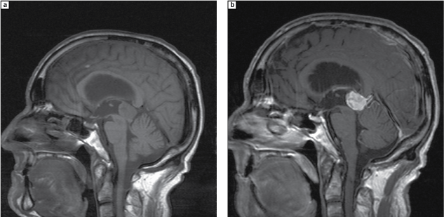
Figure 3 From Low Grade Oligodendroglioma Of The Pineal
Their importance is mainly in the fact that they cannot be distinguished from cystic tumors especially when large or when atypical features are present.
Pineal gland tectal plate. Pineal cysts are common usually asymptomatic and typically found incidentally. But we now know that pineal gland function is to control the sleep wake cycle circadian rhythm by. The pineal gland is a small pine cone shaped structure considered to be part of the epithalamus.
Mri atlas of the brain. Historically the pineal gland was considered to be the third eye because of its connections to the visual system. Cranial nerves are nerves that emerge directly from the brain in contrast to spinal nerves which emerge from segments of the spinal cordin humans there are traditionally twelve pairs of cranial nerves.
Human brain with cranial nerves. Only the first and the second pair emerge from the cerebrum. The remaining ten pairs emerge from the brainstem.
The epithalamus is the posterior part of the diencephalonit is located posteroinferior to the thalamus and consists of the pineal body stria medullaris and habenular trigone. This page presents a comprehensive series of labeled axial sagittal and coronal images from a normal human brain magnetic resonance imaging exam. This mri brain cross sectional anatomy tool serves as a reference atlas to guide radiologists and researchers in the accurate identification of the brain structures.

The Midbrain Chapter 4 Clinical Neuroradiology
Pineal Cyst Glial And Other Rare Tumors
High Prevalence Of Pineal Cysts In Healthy Adults
Pineal Region Mass
High Prevalence Of Pineal Cysts In Healthy Adults
:background_color(FFFFFF):format(jpeg)/images/library/7369/A5HDQYRFdSF2pSwX6AzQ_Glandula_pinealis_1.png)
Pineal Gland Anatomy Histology And Blood Supply Kenhub
:background_color(FFFFFF):format(jpeg)/images/library/1335/Habenular_trigone.png)
Habenular Nuclei Anatomy And Clinical Aspects Kenhub
Imaging And Differential Diagnosis Of Tectal Plate Gliomas
Grodesch Avms Of The Tectal Plate